3D (bio)printing of musculoskeletal tissues

3D (bio)printing of musculoskeletal tissues
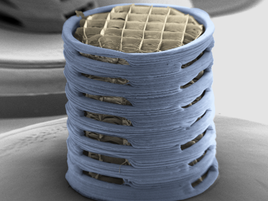
In recent years we have utilised emerging biofabrication and bioprinting strategies to engineer prevascularised implants for bone repair (https://doi.org/10.1016/j.actbio.2021.03.003) and structurally organised articular cartilage (https://doi.org/10.1016/j.biomaterials.2022.121405) and meniscus tissue (https://doi.org/10.1016/j.actbio.2022.12.047). We have also developed a range of different bioinks capable of supporting distinct cellular phenotypes, and used these bioinks to bioprint cartilage (https://onlinelibrary.wiley.com/doi/full/10.1002/adhm.201801501#) and meniscal (https://doi.org/10.1002/term.2602) grafts. As part of my ERC consolidator grant, we modified such inks for gene (10.1016/j.jconrel.2019.03.006 & 10.1089/ten.tea.2016.0498) and growth factor (https://doi.org/10.1016/j.actbio.2021.04.016) delivery, and to provide them with unique mechanical properties compatible with load bearing environments (10.1088/1758-5090/ab8708 & 10.1088/1758-5090/ac41a0). Furthermore, we bioprinted implants containing spatiotemporally defined patterns of growth factors and demonstrated that printed constructs containing a gradient of VEGF, coupled with spatially defined BMP-2 localization and release kinetics, accelerated large bone defect healing with little heterotopic bone formation (https://doi.org/10.1126/sciadv.abb5093). We have also used 3D printing techniques to produce fibre-reinforced cartilaginous templates, and assessed the efficacy of such constructs in a caprine model of osteochondral defect repair (https://doi.org/10.1016/j.actbio.2020.05.040). We have also demonstrated how emerging additive manufacturing techniques such as melt electrowriting (https://doi.org/10.1016/j.addma.2022.102998) can be used to produce scaffolds capable of directing angiogenesis and the regeneration of large bone defects (https://onlinelibrary.wiley.com/doi/full/10.1002/adhm.202302057#).
Example Project: https://x.com/dannykelly1978/status/1722299860290826586?s=20